
Figure 5.

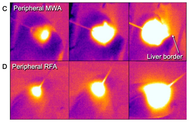
Thermography in central and peripheral ablations. From left to right: Comparative examples of a central (A) MWA and (B) RFA sequence to a peripheral (C) MWA and (D) RFA sequence. Note the adjacent vessels with a visible heat sink in both techniques but how the shape of central RFA changes compared to peripheral RFA. Further, note how heat transmission at the liver border is visualized and might help in preventing injuries at nearby structures/organs (peripheral MWA, right frame). RFA, radiofrequency ablation; MWA, microwave ablation.