Figure 1.
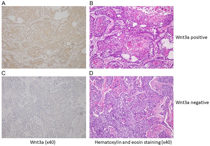
Wnt3a expression by immnohistochemical staining for resected specimen with ESCC. (A) Representative specimen with Wnt3a-positive ESCC. Cytoplasm was uniformly stained in ESCC tissue. (B) Hematoxylin and eosin (H&E) staining in ESCC tissue same as A. (C) Representative specimen with Wnt3a-negative ESCC. Cancer cells were not applicable stained at the same as stroma cells. (D) H&E staining in ESCC same as C.

