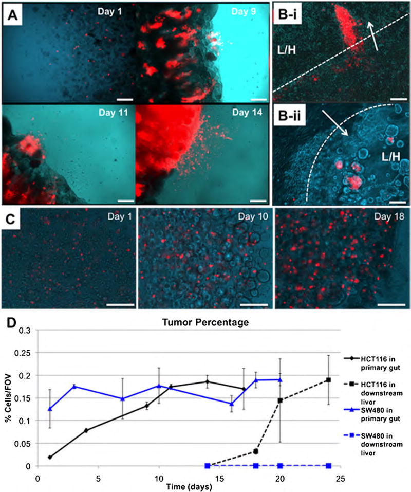
Figure 2.
HCT-116 colon carcinoma cells metastasize from the gut to the liver, while less malignant SW480 colon carcinoma cells do not. A: Growth of HCT-116 cells in the primary gut construct, and subsequent shedding of RFP-labeled HCT-116 cells into circulation. B: (i–ii) Invasion of RFP-labeled HCT116 cells into liver constructs, via multicellular protrusions and aggregates invading a liver-hydrogel construct. (Arrow—direction of invasion; Dotted line—construct interface; L/H—liver/hydrogel construct). C: SW480 cells grow in the primary construct, but never appear to shed into circulation or colonize the downstream liver construct. D: Quantification of the relative area per field of view occupied by HCT-116 cells (Black lines) and SW480 cells (Blue lines). Solid lines indicate quantification in the primary gut construct and dashed lines indicate quantification in the downstream liver construct. Scale bars—250 µm.

