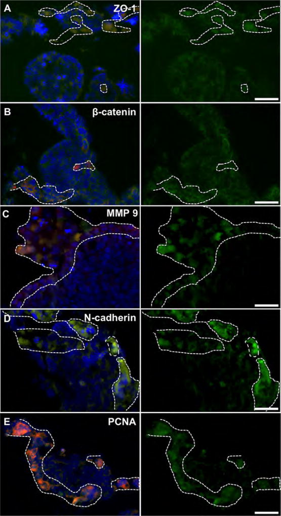
Figure 3.
Marker expression of RFP-labeled HCT-116 tumor cells (dashed line) inside 3-D intestine constructs. HCT-116 regions do not clearly express membrane-bound (A) ZO-1, or (B) β-catenin, indicating a more motile phenotype. β-catenin accumulates in the cytoplasm/nucleus suggesting WNT pathway activation, common in metastatic cells after epithelial-to-mesenchymal transition. RFP-labeled HCT-116 cells express (C) MMP-9, (D) N-cadherin, and (E) PCNA indicating a mesenchymal and metastatic proliferative phenotype. Conversely, intestine epithelial regions (no RFP) exhibit traditional epithelial phenotype with membrane-bound ZO-1, and β-catenin, while expressing MMP-9, N-cadherin and PCNA less strongly. Green—indicated stain; Red—RFP-labeled HCT-116 cells; Blue—DAPI. Scale bars—50 µm. Dotted white lines outline RFP+ HCT-116 rich regions.

