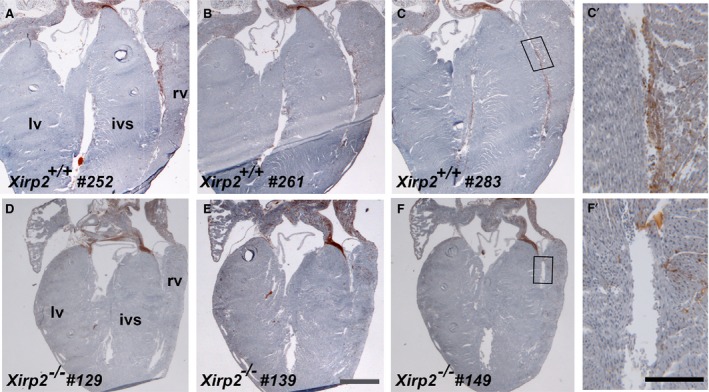
Figure 6.

Acetylcholine esterase (AChE) stain revealing a reduction in Purkinje fibers of P18 Xirp2 −/− heart. Serial 10‐μm sections (sectioning from frontal/ventral to dorsal) of entire hearts from Xirp2 +/+ and Xirp2 −/− mice were histochemically stained for AChE activity (brown) to reveal the ventricular conduction system. Purkinje fibers were positively stained in representative sections of Xirp2 +/+ heart (a, section #252; b, #261; and c, #283), whereas comparable sections of Xirp2 −/− heart showed a drastically reduced in AChE activity (d, section #129; e, #139; and f, #149). Bar in e=1 mm for a through f. rv, right ventricle; lv, left ventricle; ivs, interventricular septum. (c′ and f′) higher magnification of the box regions in c and f, respectively. Bar in f’=0.2 mm for c′ and f′.
