Figure 2.

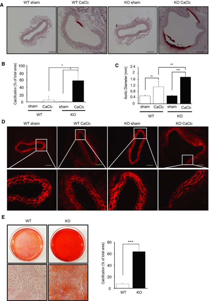
Deletion of IKKβ in smooth muscle cells promotes calcification. A, Representative microscopy images of cross sections of infrarenal aortas stained with Alizarin Red from wild‐type (WT) and IKKβ knockout (KO) littermate mice 2 weeks after saline (sham) or CaCl2 treatment. B, Quantification of vascular calcification. Calcification was quantified by ImageJ software. Graph presented is the percentage of positively stained medial layer areas in the total area of medial layer. Bars represent the mean±SD (2‐way ANOVA, n=5). C, Graph is the maximal external diameter of the WT and KO infrarenal aorta 2 weeks after saline (sham) or CaCl2 treatment. Bars represent the mean±SD (2‐way ANOVA, n=5). D, Representative fluorescence microscopy images of medial elastic lamellae in the cross sections of infrarenal aortas 2 weeks after saline (sham) or CaCl2 treatment. E, Representative microscopy images of Alizarin Red staining of 4‐week cultured vascular smooth muscle cells (VSMCs) isolated from WT and KO littermate mice. Calcification was quantified by ImageJ software. Graph presented is the percentage of positively stained area in the total area randomly selected. VSMCs were cultured in normal medium with 10% fetal bovine serum. Bars represent the mean±SD (t test, n=6). *P<0.05, **P<0.01, ***P<0.001.
