Figure 4.

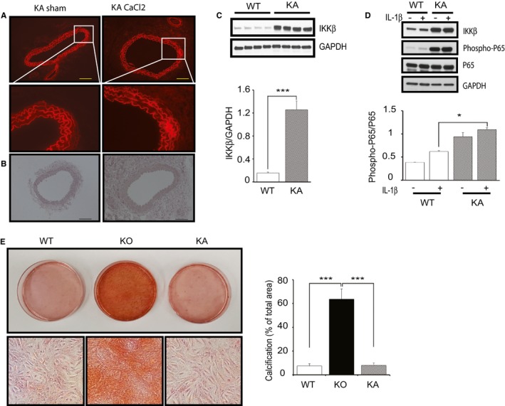
Constitutive activation of IKKβ in vascular smooth muscle cells (VSMCs) inhibits calcification. A, Representative fluorescence microscopy images of medial elastic lamellae in the cross sections of infrarenal renal aortas from kinase‐active IKKβ (KA) mice 2 weeks after saline (sham) or CaCl2 treatment. Bar=50 μm. B, Representative microscopy images of cross sections of infrarenal aortas stained with Alizarin Red from KA mice 2 weeks after saline (sham) or CaCl2 treatment. Bar=50 μm. C, Representative Western blots and densitometric analysis of IKKβ/GAPDH expression in cultured VSMCs isolated from wild‐type (WT) and KA littermate mice. Bars represent the mean±SD (t test, n=4). D, Representative Western blots and densitometric analysis of phosphorylated p65/total p65 expression in cultured VSMCs isolated from WT and KA littermate mice with stimulation by interleukin (IL)‐1β (2.5 ng/mL) or the vehicle for 15 minutes. Bars represent the mean±SD (2‐way ANOVA, n=3). E, Representative microscopy images of Alizarin Red staining of 4‐week cultured VSMCs isolated from WT, IKKβ knockout (KO), and KA mice and quantification of VSMC calcification. VSMCs were cultured in normal medium with 10% fetal bovine serum. Calcification was quantified by ImageJ software. Graph presented is the percentage of positively stained area in the total area randomly selected. Bars represent the mean±SD (1‐way anova, n=6). *P<0.05, ***P<0.001.
