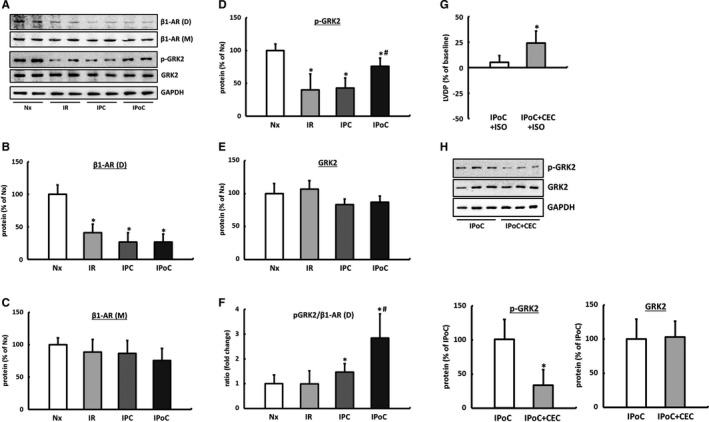
Figure 3.

Influence of IR, IPC, and IPoC on β‐adrenergic signaling 2 hours after starting reperfusion. A, Representative immunoblots and quantification by densitometric analysis of (B) β1‐AR dimers (D, MW 95 kDa) and (C) β1‐AR monomers (M, MW 50 kDa). Quantification of the (D) phosphorylated (p‐GRK2) and (E) total GRK2 protein. F, The p‐GRK2 to β1‐AR (D) ratio was calculated for each animal, and group means were expressed as fold change of Nx. Data are means±SD of n=6 hearts. *P≤0.05 vs Nx, # P≤0.05 vs IPC. The significance of PKC for GRK2 phosphorylation. G, ISO‐induced inotropic response of IPoC hearts perfused with the PKC inhibitor CEC. H, The effect of PKC inhibition on GRK2 phosphorylation in IPoC hearts. Immunoblot bands were normalized to GAPDH and expressed as a percentage of Nx. Data are means±SD of n=6 hearts. *P≤0.05 vs IPoC. CEC indicates chelerythrine chloride; D, dimer; GAPDH, Glyceraldehyde 3‐phosphate dehydrogenase; GRK2, G protein‐coupled receptor kinase 2; IPC, ischemic preconditioning; IPoC, ischemic postconditioning; IR, ischemia/reperfusion; ISO, isoproterenol; M, monomer; Nx, normoxic; p‐GRK2, phosphorylated GRK2; PKC, protein kinase C; β1‐AR, β1‐adrenergic receptor.
