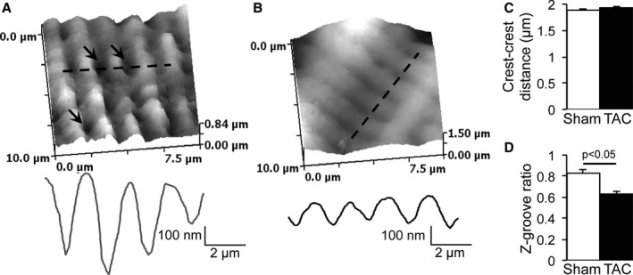
Figure 4.

Structural remodeling of the lateral membrane of transverse aortic constriction (TAC) cardiomyocytes. A, Typical scanning ion conductance microscopy (SICM) scan and topology profile along the dashed line of a sham–operated on (Sham) cardiomyocyte lateral membrane (arrows indicate T‐tubule openings). B, Typical SICM scan and topology profile along the dashed line of a TAC cardiomyocyte lateral membrane. C, Unchanged crest‐crest distance between Sham and TAC. D, Decreased Z‐groove ratio in TAC compared with Sham.
