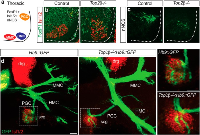
Figure 6.
Defects in PGC columnar identities and peripheral projections in Top2β-/- mice. A, Organization of motor columns at thoracic levels of the spinal cord; PGC, preganglionic motor column, MMC, medial motor column, HMC, hypaxial motor column. B, C, Expression of PGC markers FoxP1 (white circle; B) and nNOS (C) is dramatically reduced in Top2β-/- embryos. The remaining neurons expressing nNOS are displaced ventrally. Scale bar = 50 μm. D, PGC neurons project to scg in Top2β-/-mice but show an aberrant innervation pattern at the target. scg: sympathetic chain ganglia and drg: dorsal root ganglia. Scale bar = 50 μm.

