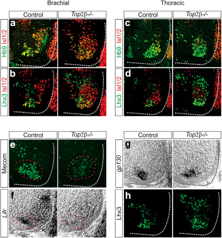
Figure 9.

Defects in MN organization and MMC specification in Top2β-/- mice. A–D, Disorganization of MNs in Top2β-/- mice at e12.5 at brachial (A, B) and thoracic (C, D) levels of the spinal cord. Hb9+ and Isl1/2+ MNs do not appear to segregate in Top2β-/- mice, similar to the phenotype observed in Pbx1/3 mutant animals. Scale bar = 50 μm. E, F, Effects of Top2β deletion on the expression of MMC molecular markers at brachial levels of the spinal cord at e12.5. Mecom and Lifr (red circles), both targets of Pbx proteins, are downregulated in Top2β-/-mice. G, H, Expression of gp130 (G) and Lhx3 (H) persist in Top2β-/-mice at e12.5. Dorsal Lhx3 expression corresponds to a population of V2 interneurons that appears to be expanded in Top2β-/- mice.
