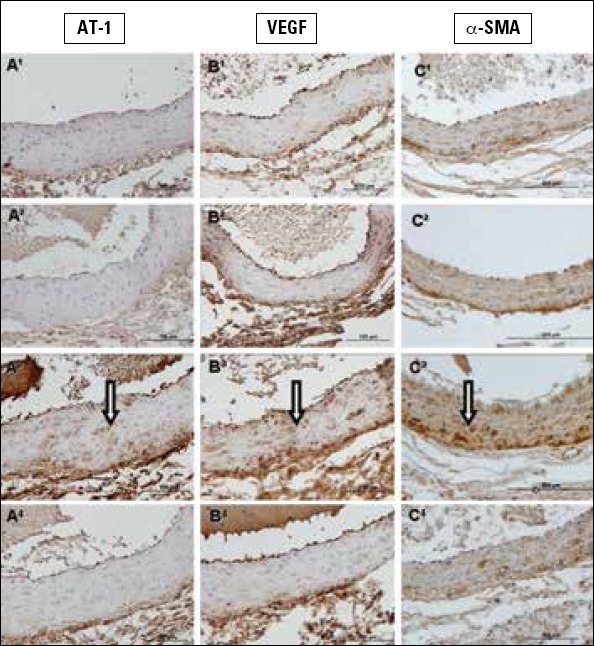
Figure 3.

1A-C. Sections belonging to the control group, 2A-C. sections belonging to the LA group, 3A-C. sections belonging to the 5/6 Nx group, and 4A-C. 5/6 Nx + LAT groups. Arrows show immunopositive aorta tissue. A1-4. Stained with type I angiotensin (AT1) receptor, B1-4. stained with vascular endothelial growth factor, and C1-4. stained with alpha-smooth muscle actin immunohistochemically. Scale bar 200 μm
