. 2017 Sep 6;6:e27891. doi: 10.7554/eLife.27891

Figure 1. A complex containing eIF4E, Cup, TRAL, ME31B, and PABP is abundant in 0–1 hr embryos.

(A) Selected results from mass spectrometry of PABP and eIF4G co-immunoprecipitations in S2 cells. Extracts from S2 cells were immunoprecipitated with Fab C1 (a negative control), anti-PABP or anti-eIF4G Fabs. Shown are the peptide counts and protein size for proteins of interest. (B) As in (A), but for extracts from 0 to 1 hr embryos. (C) Western blot analysis for proteins co-immunoprecipitated with PABP and eIF4G in S2 cells. Western blots of input and immunoprecipitates were probed for the indicated proteins. (D) As in (C), but for extracts from 0 to 1 hr embryos. (E) Proteins binding m7GDP beads in S2 cell extracts. Lysates were incubated with either blank agarose beads or beads conjugated with m7GDP. Western blots of the input and bound fractions are shown for the indicated proteins. (F) As in (E), except with 0–1 hr embryo extracts.

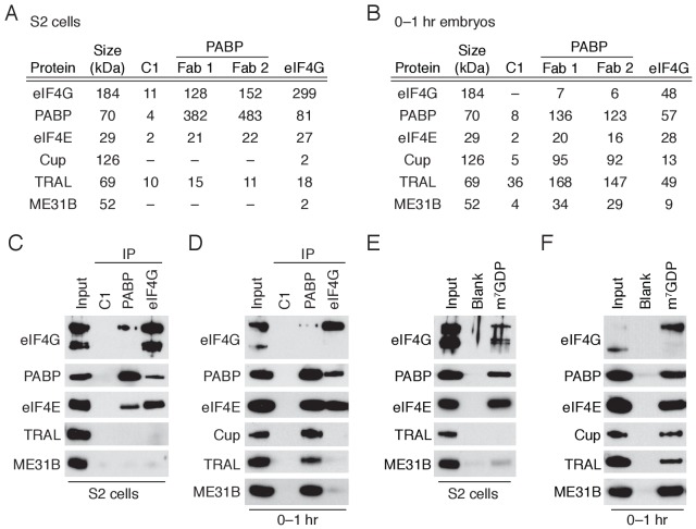
Figure 1—source data 1. Mass spectrometry results for PABP and eIF4G immunoprecipitations in S2 cells.
DOI: 10.7554/eLife.27891.004
Figure 1—source data 2. Mass spectrometry results for PABP and eIF4G immunoprecipitations in 0–1 hr embryos.
DOI: 10.7554/eLife.27891.005

Figure 1.

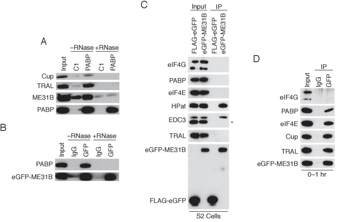
Figure 1—figure supplement 1. ME31B interacts with PABP and eIF4E in the early embryo.

Figure 1—figure supplement 1.

(A) RNA-dependence for the interactions between PABP and ME31B, Cup, and TRAL. 0–1 hr embryo extracts were co-immunoprecipitated with either the control or anti-PABP Fabs in the presence or absence of RNase A. Western blots of inputs and immunoprecipitates are shown for the indicated proteins. (B) As in (A), except for ME31B immunoprecipitation. 0–1 hr eGFP-ME31B embryo extracts were co-immunoprecipitated with either rabbit IgG or anti-GFP antibodies in the presence or absence of RNase A. (C) Western blot analysis for proteins interacting with ME31B in S2 cells. Cells were transfected with either FLAG-eGFP or eGFP-ME31B, and then co-immunoprecipitation was performed with anti-GFP antibodies. Western blots of input and immunoprecipitates are shown for the indicated proteins; in the lower panel, proteins were probed with anti-GFP. The arrowhead indicates a nonspecific band. (D) Western blot analysis for proteins interacting with ME31B in 0–1 hr embryos. Extracts from 0 to 1 hr eGFP-ME31B embryos were co-immunoprecipitated with anti-GFP or rabbit IgG. Western blots of input and immunoprecipitates are shown for the indicated proteins.