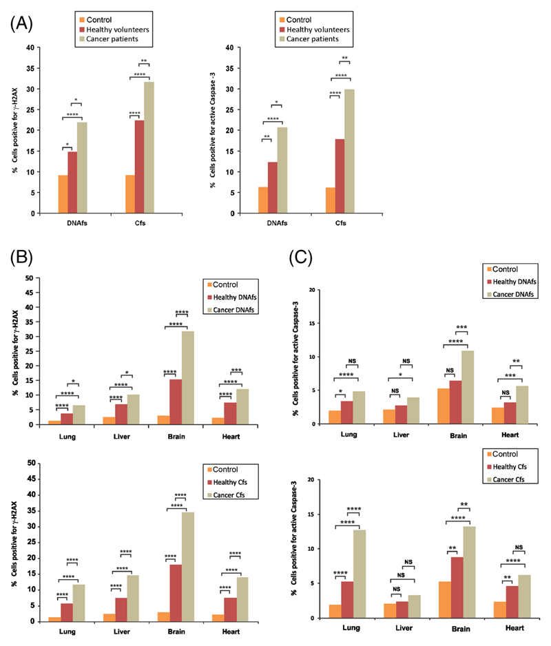
Figure 4.
Induction of γH2AX and active Caspase-3 by DNAfs and Cfs derived from healthy volunteers. Samples from cancer patients were also analysed for comparison. DNAfs and Cfs were isolated from pooled plasma/serum of healthy volunteers and age- and sex-matched cancer patients. (A) In vitro analysis of γ-H2AX and active Caspase-3. NIH3T3 cells (10×104) were treated with DNAfs and Cfs (5 ng DNA each) for 6 h for detection of γ-H2AX (left) and for 24 h for detection of active Caspase -3 (right) by immuno-fluorescence. For γ-H2AX (left-hand panel), 300 nuclei were counted and the percentage of nuclei showing positive foci were calculated and analysed by Chi-square test. For active Caspase-3 (right-hand panel), 200 cells were counted and the percentage of cells showing positive fluorescent signals were calculated and analysed by Chi-square test. *p<0.05, **p<0.01, ****p<0.0001. (B) In vivo detection of γ-H2AX activation by DNAfs (upper panel) and Cfs (lower panel). Mice were injected intravenously with DNAfs and Cfs (100 ng DNA each) and vital organs were removed after 24 h and processed for γH2AX by immuno-fluorescence as described earlier. Control animals were injected with 100 μl of saline. The experiments were done in duplicate i.e., with two animals in each group. One thousand cells from each animal were analysed and the percentage of nuclei with positive fluorescent foci was calculated and analysed by Chi-square test. *p<0.05, **p<0.01, ****p<0.0001. (C) In vivo analysis of Caspase-3 activation by DNAfs (upper panel) and Cfs (lower panel). Mice were injected intravenously with DNAfs and Cfs (100 ng DNA each) and vital organs were removed after 24 h and processed for active Caspase-3 by immuno-fluorescence as described earlier. Control animals were injected with 100 μl of saline. The experiments were done in duplicate i.e., with two animals in each group; one thousand cells from each animal were analysed and the percentage of cells with positive fluorescence was calculated and analysed by Chi-square test. *p<0.05, **p<0.01, ***p<0.001, ****p<0.0001.

