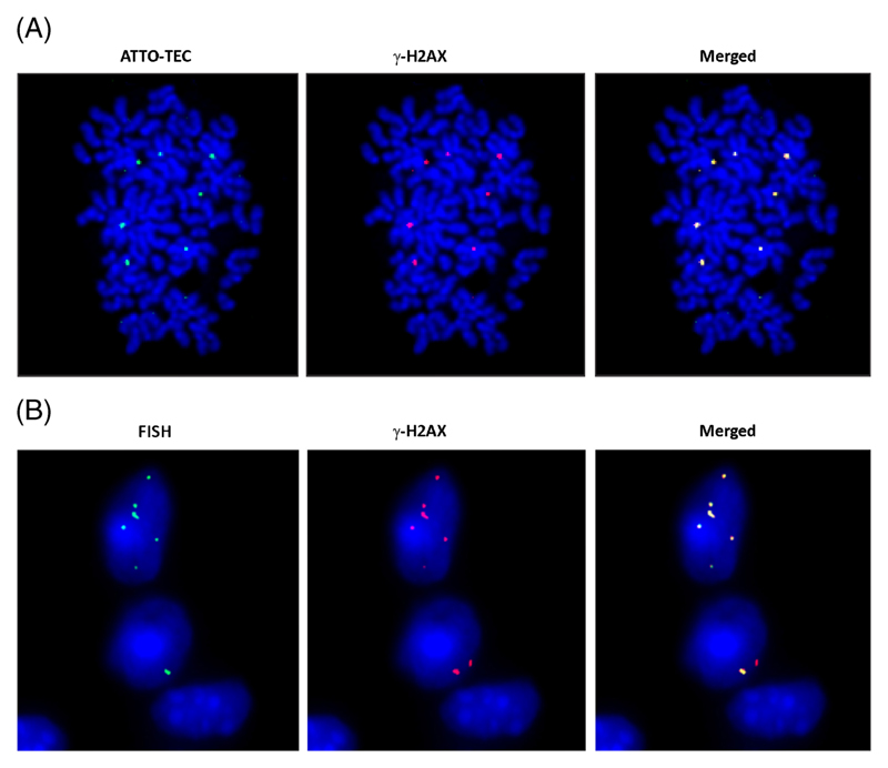
Figure 5.
Genomic integration of Cfs involves DNA double-strand break repair. (A) In vitro demonstration of γ-H2AX foci at sites of Cfs integration. NIH3T3 cells (10×104) were treated for 6 h with Cfs (5 ng DNA) labelled in their protein with ATTO-TEC (green) and metaphase spreads were prepared. Immuno-fluorescence images were developed using antibody to γ-H2AX (red). Co-localization of green and red signals were clearly visible. (B) In vivo demonstration of γ-H2AX foci at the sites of Cfs integration. Mice were injected i.v. with Cfs (100 ng DNA) and sacrificed 24 h later. Sections of brain were processed for immuno-FISH using human-specific genomic probe (green) and antibody against γ-H2AX (red). Co-localization of green and red signals are clearly visible.

