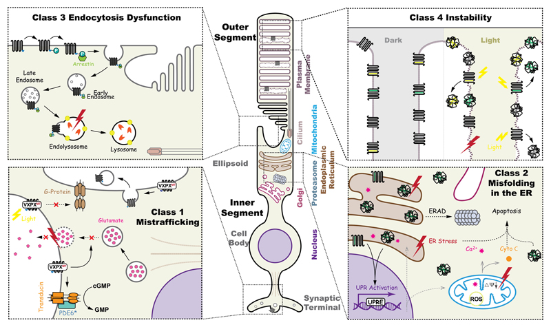
Figure 3. Schematic illustration of the potential pathogenic consequences caused by rhodopsin mutations.
A rod photoreceptor has distinct regions; including, the outer segments (OS) and inner segments (IS), cell body and synaptic terminals. Pathogenic mutations in rhodopsin disturb several cellular pathways, including endocytosis dysfunction, structural instability of the OS, mistrafficking, and misfolding. Top left, class 3, hyperphosyphorylated rhodopsin is bound by arrestin (green), the Rho-arrestin complex is endocytosed and disrupts vesicular traffic. Top right, upon illumination, unstable rhodopsin mutants (class 2, 4 and 7) could aggregate in the disk of OS, thus causing damage of the plasma membrane homeostasis. Bottom left, class 1 mutations that affect the ciliary targeting signal, VXPX, are mislocalised, including at the synapse, where they could inhibit synaptic vesicle fusion or be abnormally activate transducin. Misfolding class 2 rhodopsin mutants in the ER might induce ER stress, degraded by ERAD and potentially aggregate, leading to the activation of pro-apoptotic pathways.

