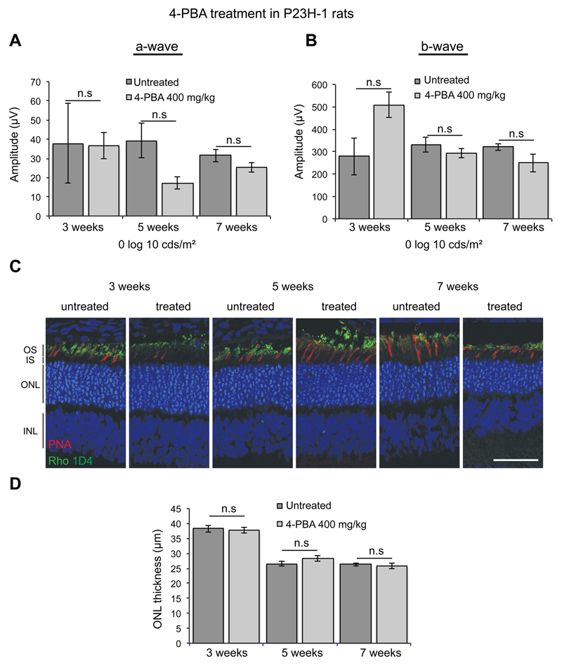
Figure 4. 4-PBA does not affect photoreceptor function and survival in P23H-1 rats.
P23H-1 rats were either left untreated or treated from P21 to P49 with 400 mg/kg 4-PBA (n = 4 for each condition) via IP injection. (A) Scotopic ERG’s were performed as previously described (Aguila et al., 2014). No significant differences were observed in P23H-1 rats that had received the treatment at any time point and at any intensity (-6 to 2.7 log cds/m2) (data not shown). Average at 0 log cds/m2 for a-wave and b-wave of P23H-1 rats at 3, 5 and 7 weeks of age. Values are means ± SEM. n.s non-significant values, 2 way ANOVA. (B) Retinal histology for measuring the outer nuclear layer (ONL) thickness was made on digital images of stained cryosections, every 500 microns from the optic nerve outwards for both the inferior and superior hemisphere. The ONL thickness across the whole from 4 animals at each time point was averaged and analysed using Graphpad Prism (Sigmastat). (C) Representative images of the ONL in treated and untreated P23H-1 rats at 3. 5 and 7 weeks stained with DAPI (blue), anti-rhodopsin antibody 1D4 (green) and cone marker peanut agglutinin (PNA) (red). Scale bar = 50 microns.

