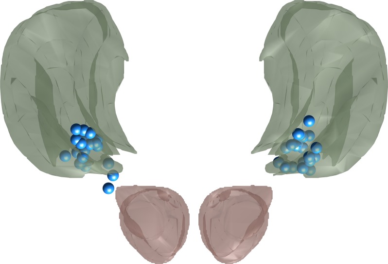
Fig 1. Projection of all active contacts (blue) onto a neuroanatomical atlas [19].
Green: VIM, red: red nucleus. Note that most active contacts lay inside the VIM proper or at its ventral border while two contacts lay slightly more ventral in the zona incerta.

