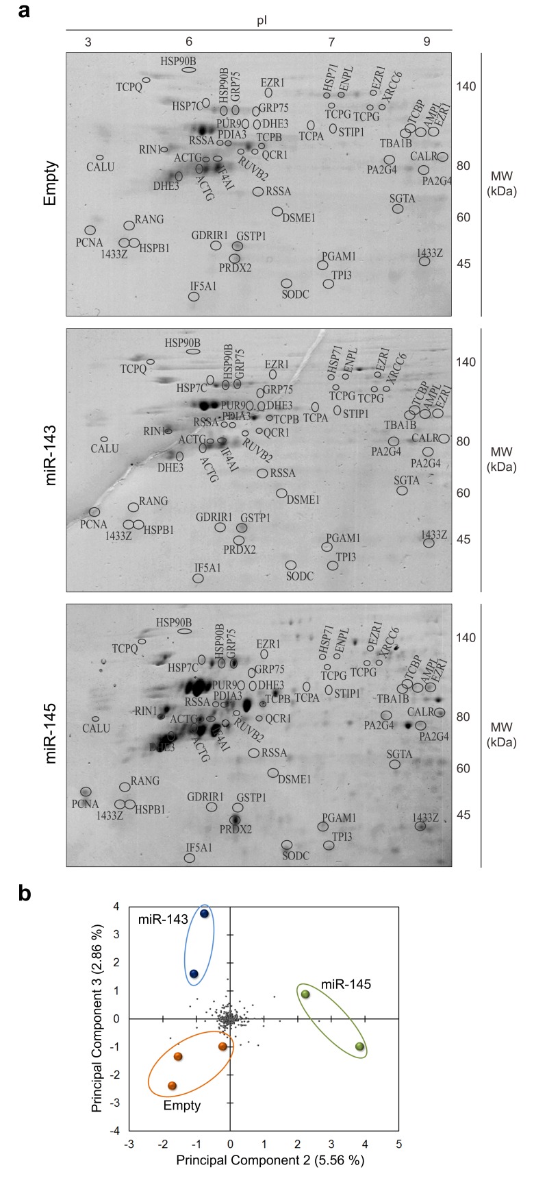
Fig 1. Two-dimensional gel electrophoresis analysis of HCT116 human colon cancer cells overexpressing miR-143, miR-145, or Empty vector.
Proteins of HCT116 stably transduced cells were separated by 2-DE (IEF (pI 3–10 non-linear) + SDS-PAGE) and visualized by staining with Coomasie brilliant blue R-350. (a) Representative 2-DE gels. Identified proteins are listed in S1 and S2 Tables. (b) Principal Component Analysis (PCA). Coloured spots represent each gel prepared for the indicated condition. Grey spots represent proteins detected in the 2-DE gels.

