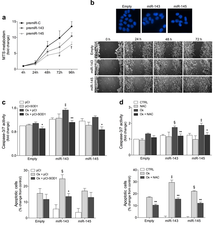
Fig 5. miR-143 and miR-145 impact on cell proliferation and morphology and miR-143-induced oxidative stress contributes to oxaliplatin-mediated apoptosis in HCT116 human colon cancer cells.
(a) The growth profiles of HCT116 cells transiently expressing miR-143 (premiR-143), miR-145 (premiR-145), or control (premiR-C) precursors were monitored by MTS metabolism assay at 4, 24, 48, 72 and 96 h after plating. (b) Nuclear morphology was evaluated by fluorescence microscopy after Hoechst staining. Representative images of Hoechst staining at 400x magnification are presented (top). Cell migration was assessed by wound healing assay at 24, 48 and 72 h after wound formation. Representative images are presented (bottom). HCT116 cells stably transduced with miR-143, miR-145, and Empty vector were treated with oxaliplatin (Ox) and transfected with pCI-neo (pCI) or pCI-neo-SOD1 (pCI-SOD1) plasmids (c), or exposed to NAC (d). Caspase-3/7 activity was determined by Caspase-Glo 3/7 assay. Apoptosis was quantified by flow cytometry using Guava Nexin assay. Results are expressed as mean ± SEM fold change and percentage change of apoptotic cells ± SEM, from at least three independent experiments. #p < 0.05 and †p < 0.01 from control precursor (premiR-C) transfected cells; ‡p < 0.01; §p < 0.05 from Empty cells treated with oxaliplatin; ***p < 0.001, **p < 0.01, *p < 0.05 from respective oxaliplatin treated cells.

