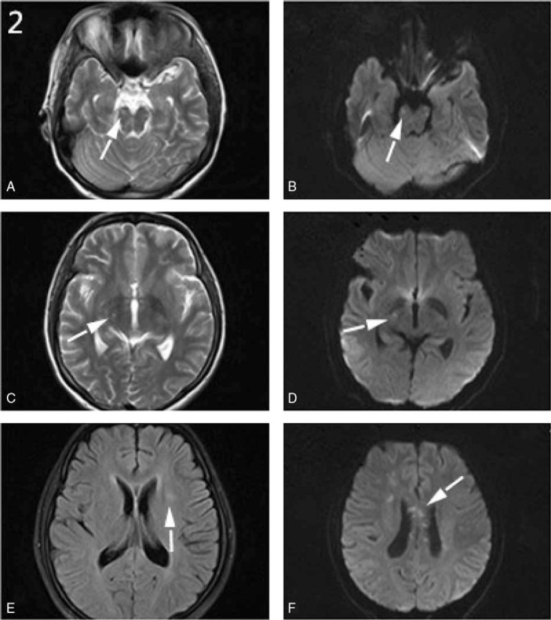
Figure 2.

A–F, Brain MRI scans of case 1 showed multiple hyperintensity lesions (A–D) in the right thalamus and pons on the T2 and DWI sequences. High-intensity lesions were also found in the left basal ganglia on the FLAIR sequence (E) as well as in corona radiate and corpus callosum on the DWI sequence (F). DWI = diffusion-weighted imaging, FLAIR = fluid-attenuated inversion recovery, MRI = magnetic resonance imaging.
