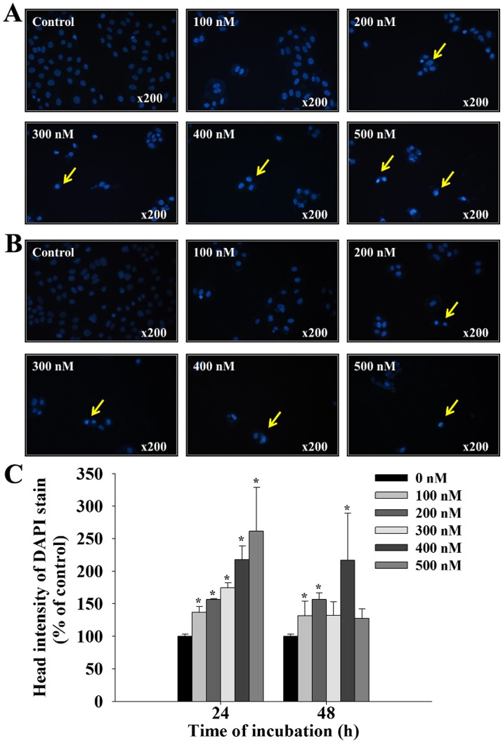
Figure 2.
Bufalin induced chromatin condensation in SCC-4 cells. Cells were treated with 0, 100, 200, 300, 400 and 500 nM of bufalin for (A) 24 and (B) 48 h and then stained with DAPI. Yellow arrows indicate areas of positive DAPI staining. (C) The intensity of DAPI staining was calculated to determine the level of chromatin condensation. The results are presented as the mean ± standard deviation (n=3). *P<0.05 vs. 0 nM bufalin (control). DAPI, 4′,6-diamidino-2-phenylindole, dilactate.

