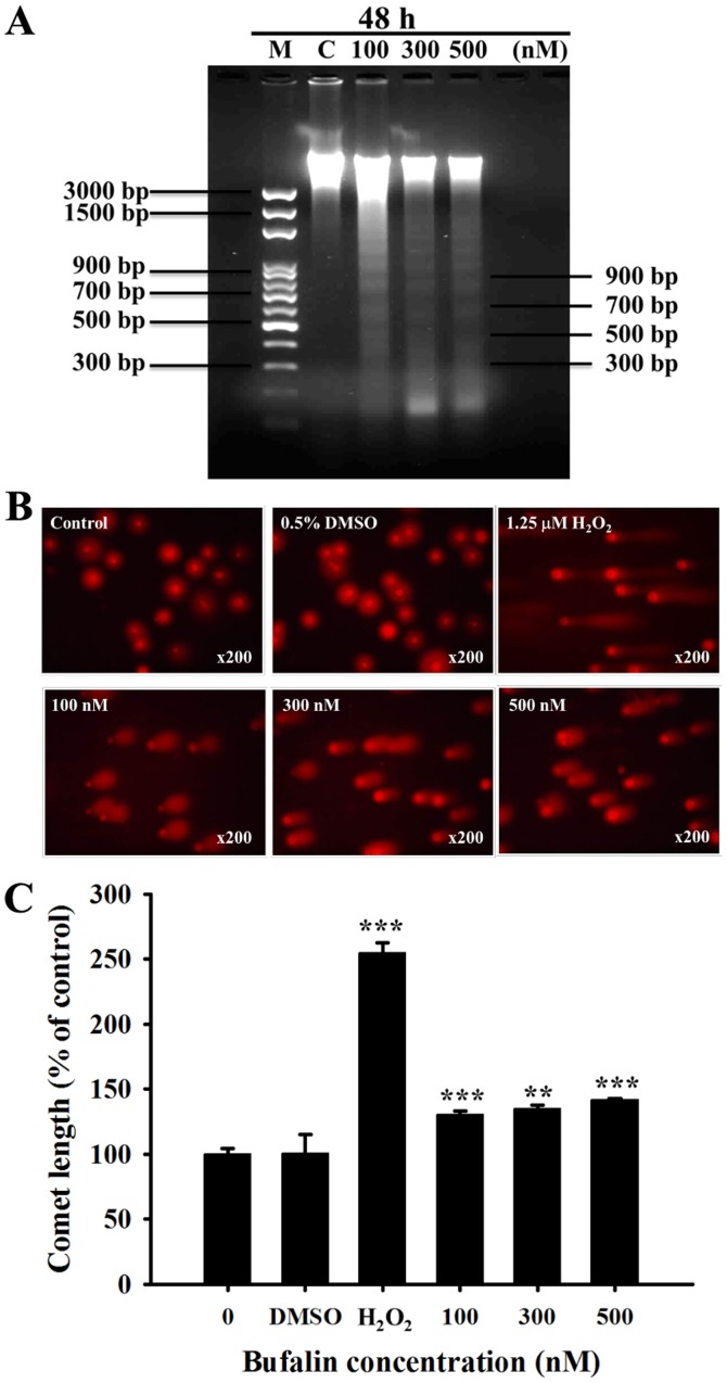
Figure 3.
Bufalin induced DNA fragmentation and damage in SCC-4 cells. Cells were treated with 0.5% DMSO, 1.25 µM H2O2, or 0, 100, 300 and 500 µM of bufalin for 48 h, and then DNA was isolated to analyze (A) DNA fragmentation (DNA ladder) by DNA gel electrophoresis or (B) to analyze DNA damage by a Comet assay and (C) quantitating the length of comet tails. Magnification, ×200. The results are presented as the mean ± standard deviation (n=3). **P<0.01 and ***P<0.001 vs. 0 nM bufalin (control). M, marker; C, control (0 nM bufalin); DMSO, dimethyl sulfoxide.

