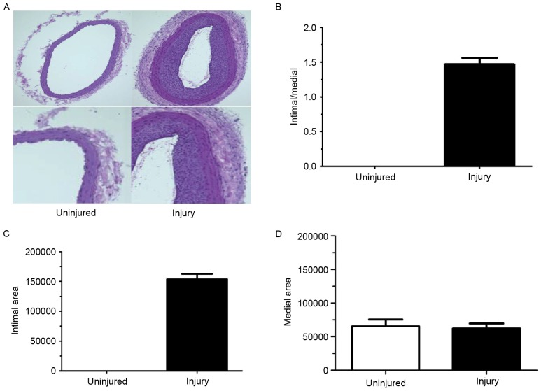
Figure 1.
Neointimal hyperplasia formation following balloon injury. (A) Tissues were evaluated by hematoxylin and eosin staining (magnification, ×200) at 28 days subsequent to balloon injury. The (B) ratio of intima/media (normalized to the uninjured tissue), (C) area of the intima and (D) area of the media were calculated and between the right and left carotid artery in the balloon injury group. Data are presented as the mean ± standard error of the mean.

