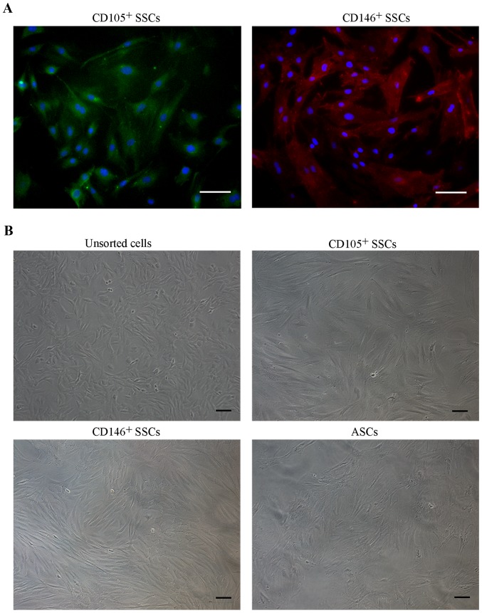
Figure 2.
Cell morphology of isolated stem cells. (A) Immunofluorescence staining was performed on CD105+ SSCs and CD146+ SSCs, the nuclei were counterstained with DAPI. (B) Morphology of unsorted growth plate cells, CD105+ SSCs, CD146+ SSCs and ASCs. Scar bar, 100 µm. ASCs, adipose-derived stem cells; SSCs, skeletal stem cells; CD, cluster of differentiation.

