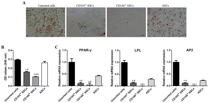
Figure 4.
Comparison of adipogenic differentiation potential. (A) Oil Red O staining of unsorted cells, CD105+ SSCs, CD146+ SSCs and ASCs. Scar bar, 200 µm. (B) Quantitative measurement of lipid droplets in all four groups. (C) The mRNA levels of PPAR-γ, LPL and AP2 were detected using reverse transcription-quantitative polymerase chain reaction. All data are from 3 independent experiments and are presented as means ± SD. *P<0.05 vs. unsorted cells; #P<0.05 vs. ASCs; αP<0.05 vs. CD105+ SSCs. PPAR-γ, peroxisome proliferators-activated receptor-γ; LPL, lipoprteinlipase; AP2, adipocyte fatty acid-binding protein 2; ASCs, adipose-derived stem cells; SSCs, skeletal stem cells; CD, cluster of differentiation.

