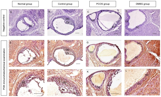
Figure 4.
Immunohistochemical examination of PI3K expression in the ovaries of each group. Negative control staining with serum instead of PI3K primary antibody in (A) blank control, (B) vehicle control, (C) PCOS and (D) DMBG groups. PI3K immunohistochemical signals appear brown and the counterstaining background appears blue in (E) blank control, (F) vehicle control (G) PCOS and (H) DMBG groups. Higher magnification images of PI3K staining are presented for (I) blank control, (J) vehicle control, (K) PCOS and (L) DMBG groups. Scale bar, 100 µm. PI3K, phosphatidylinositol 3-kinase; PCOS, polycystic ovary syndrome; DMBG, dimethyldiguanide; GC, granulosa cell; Oo, oocyte.

