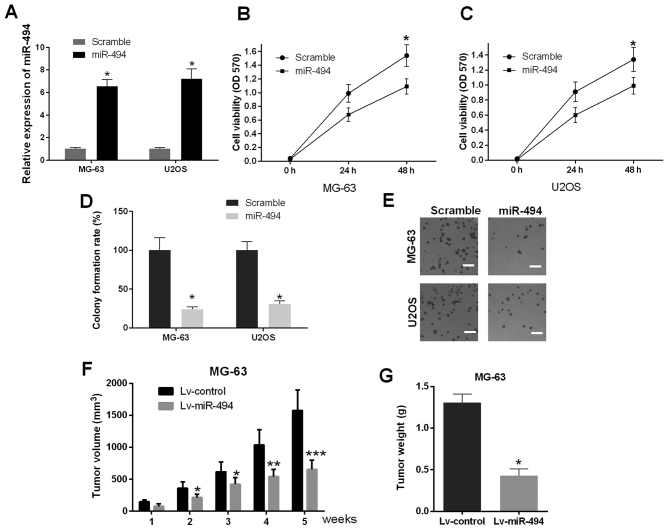
Figure 2.
miR-494 suppresses cell proliferation in vitro and in vivo. (A) Confirmation of the expression of miR-494 in MG-63 and U2OS cells transfected with miR-494 mimics and scramble. Viability of MG-63 and U2OS cells were detected using MTT assay. Overexpression of miR-494 in (B) MG-63 cells and (C) U2OS cells significantly inhibited cell viability. (D) Colony formation assay to investigate the effect of miR-494 on cell growth of MG-63 and U2OS cells. Overexpression of miR-494 in MG-63 and U2OS cells result in inhibited cell growth. (E) Representative images of MG-63 and U2OS cells under a microscope (magnification, ×200). (F) Tumor volumes were measured 1, 2, 3, 4 and 5 weeks following injection of Lv-miR-494 and Lv-control. The volumes of tumors injected with Lv-miR-494 were significantly decreased compared with those injected with Lv-control. (G) Overexpression of miR-494 significantly inhibited tumor weight in vivo 5 weeks following injection. *P<0.05, **P<0.01 and ***P<0.001. miR, microRNA; OD, optical density.

