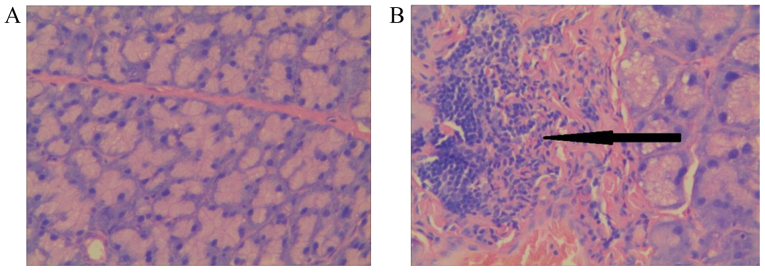
Figure 6.
Lacrimal gland histopathology (H&E staining, ×400). (A) Lacrimal gland from the Con group; (B) Lacrimal gland from the Fin group. A large number of lymphocytes had infiltrated the interlobular space and surrounded the acinar and ductal cells as indicated by the black arrow. H&E, hematoxylin and eosin.

