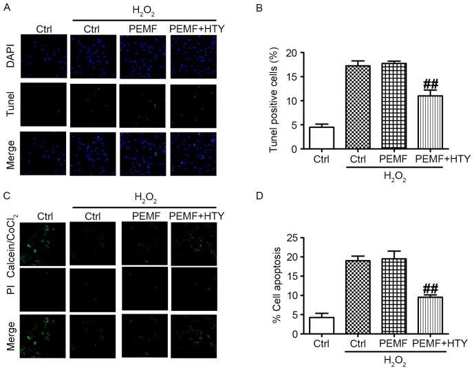
Figure 3.
Effect of HTY and PEMF on cell apoptosis under H2O2 stimulation in human umbilical vein endothelial cells. (A) Representative fluorescence images and (B) quantification of TUNEL-positive cells. DAPI staining represents the nucleus. (C) Representative fluorescence images and (D) quantification of the ratio of apoptotic cells at 24 h, as assessed by calcein acetoxymethyl/PI staining. Data are expressed as the mean ± standard error of six independent experiments. ##P<0.01 vs. PEMF-treated group. Magnification, ×200. HTY, hydroxytyrosol; PEMFs, pulse electromagnetic fields; Ctrl, control; PI, propidium iodide; TUNEL, terminal deoxynucleotidyl transferase dUTP nick-end labeling.

