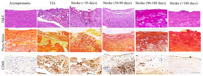
Figure 1.
Histological characteristics of plaques. Representative images of plaques in asymptomatic, TIA, stroke patients of <30, 30–90, 90–180 and >180 days. H&E staining showed inflammatory cell infiltrations (TIA, Stroke <30 d and Stroke 30–90 d groups) and intraplaque hemorrhage (TIA, Stroke <30 d groups) in the vulnerable plaques and intact cap and predominantly fibrous tissue in the stable plaques (Asymptomatic, Stroke 90–180 d and Stroke >180 d groups). Picro-Sirius Red staining showed the large cholesterol crystals in the lipid core in vulnerable plaques (TIA, Stroke <30 d and Stroke 30–90 d groups) and collagen fibrous tissue in stable plaques (Asymptomatic, Stroke 90–180 d and Stroke >180 d groups). CD 68 staining showed histological appearance of a plaque with substantial macrophage infiltration in the vulnerable plaques (TIA, Stroke <30 d and Stroke 30–90 d groups) and minor macrophage infiltration in stable plaques (Asymptomatic, Stroke 90–180 d and Stroke >180 d groups). Scale bar: 100 µm.

