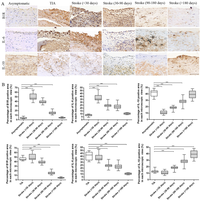
Figure 3.
Immunohistochemical results. (A) Representative immunohistochemical images of B1R, IL-6 and IL-10 in plaques of asymptomatic, TIA and stroke patients of <30, 30–90, 90–180 and >180 days. (B) Quantification of immunohistochemistry analysis of B1R, IL-6 and IL-10 staining of section from asymptomatic, TIA and stroke patients of <30, 30–90, 90–180 and >180 days. Percentage of positive staining area in each microscopic view (at 200x) showed that stroke plaques experienced initial increases of B1R and IL-6 contents at <30 days, and subsequent reduction from 30–90 to >180 days. However, a noticeable increase in IL-10 contents was found in stroke plaques from <30 to >180 days. (***P<0.001 vs. asymptomatic or TIA group). Scale bar: 100 µm.

