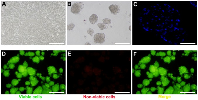
Figure 1.
2D and 3D culture of mouse BM-MSCs. (A) BM-MSCs in traditional monolayer culture. (B) Formation of spheroids following 24 h of rocking culture. (C) DAPI staining of spheroids. (D) Green staining of viable spheroids, (E) red staining of non-viable spheroids and (F) merged image of viable and non-viable spheroids, as determined by FluoroQuench™ staining. Scale bars, 200 µm. BM-MSCs, bone marrow-derived mesenchymal stem cells.

