Figure 3.
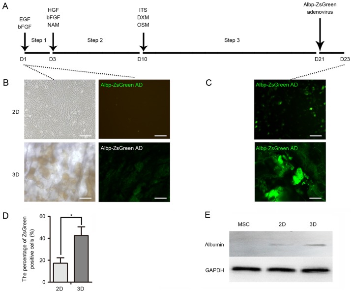
Hepatocyte-like cell induction. (A) Timeline of hepatic induction of mouse MSCs for 2D and 3D cultures. Cells were incubated in medium containing various growth factors for 23 days. Albp-ZsGreen adenovirus was added to induce expression of ALB. (B) Green fluorescence of Albp-ZsGreen adenovirus indicated ALB synthesis in non-hepatic or (C) hepatic differentiated MSCs. (D) Percentage of Albp-ZsGreen-positive hepatocyte-like cells in each group. (E) Western blot analysis demonstrated the expression of ALB in undifferentiated MSCs and following hepatic differentiation using 2D or 3D models. Data represent the mean ± standard deviation of three independent experiments. *P<0.05. Scale bars, 100 µm. MSCs, mesenchymal stem cells; ALB, albumin; EGF, epidermal growth factor; bFGF, basic fibroblast growth factor; HGF, hepatocyte growth factor; NAM, nicotinamide; DXM, dextromethorphan; OSM, oncostatin M; ITS, insulin transferrin selenium.

