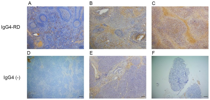
Figure 1.
Expression of IgG4 in IgG4-RD and IgG4-negative controls. (A) A large number of IgG4-positive cells infiltrated the lymph node of an IgG4-RL sample. (B) Pancreas sample from type 1 autoimmune pancreatitis. (C) Lacrimal gland sample from an IgG4-ROD patient. (D) Lymph node sample from the IgG4-negative control exhibiting no IgG4-positive cells. (E) Pancreatic specimen from the IgG4-negative control exhibiting no IgG4-positive cells. (F) Orbital tissue from the from the IgG4-negative control exhibiting no IgG4-positive cells. Scale bar, 200 µm; magnification, ×40. Ig, immunoglobulin; RD, related disease; RL, related lymphadenopathy; ROD, related ophthalmic disease.

