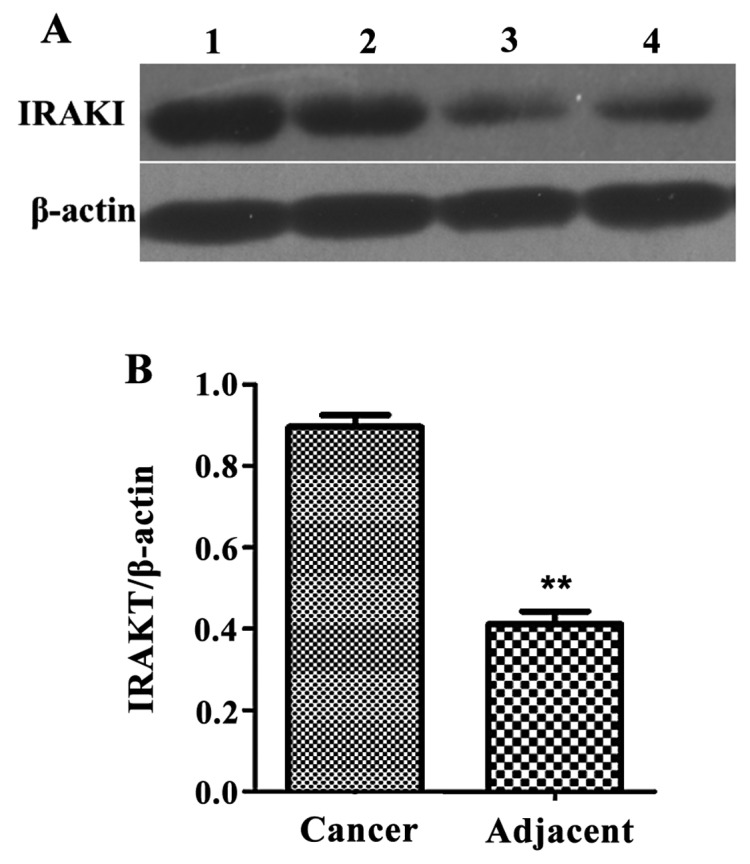
Figure 7.

Western blot detection of IRAKI protein expression levels. (A) Western blot analysis, lanes 1 and 2 show papillary thyroid carcinoma samples; lanes 3 and 4 show the coresponding adjacent tissues. (B) Relative expressions of IRAK1 protein, compared with the adjacent tissues, the expression level of IRAKI protein in the cancer tissues of papillary thyroid carcinoma was significantly decreased (**P<0.01).
