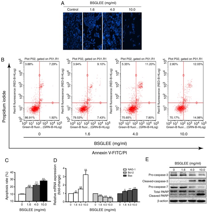
Figure 3.
Effect of BSGLEE on cell apoptosis in HCT116 cells. (A) Result from Hoechst 33342 staining. Cells were treated with different concentration of BSGLEE for 36 h. (B) Flow cytometry determination of apoptotic cells after treatment with different concentration of BSGLEE (0, 1.6, 4.0 and 10.0 mg/ml) for 36 h. Figure shows a representative image from three independent experiments. (C) Quantified histograms of the apoptosis ratio of HCT116 cells upon BSGLEE treatment. (D) Quantification of the mRNA levels of apoptosis-related genes after treatment with BSGLEE as determined qRT-PCR. (E) The expression of apoptosis-associated proteins as assessed by western blot analysis. β-actin was used as an internal control. Statistical data represent mean ± SD of three independent experiments, *P<0.05, **P<0.01.

