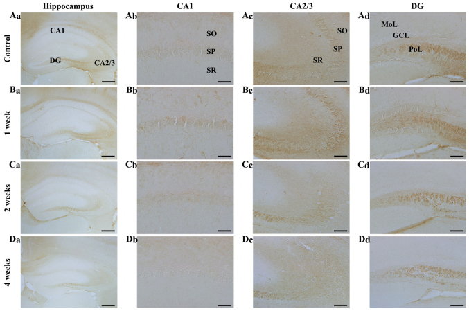
Figure 5.
NF-L immunohistochemistry in the hippocampal subregions of the (A) control and SCO-treated mice following (B) 1, (C) 2 and (D) 4 weeks. NF-L immunoreactivity was increased in the (Bc) SP 1 week following SCO treatment; however, NF-L immunoreactivity decreased in all hippocampal subregions, (C and D) 2 and 4 weeks following SCO treatment. Scale bar=800 µm (Aa-Da), 50 µm (Ab-Db) and 100 µm (Ac-Dc and Ad-Dd). CA, cornus ammonis; DG, dentate gyrus; GCL, granule cell layer; Mol, molecular layer; PoL, polymorphic layer; SO, stratum oriens; SP, stratum pyramidale; SR, stratum radiatum; NF-L, neurofilament-68 kDa; SCO, scopolamine.

