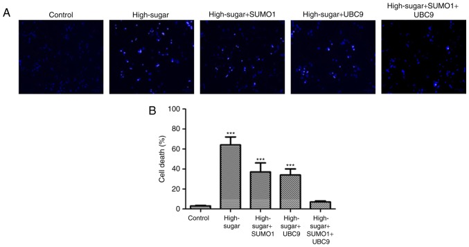
Figure 2.
Evaluation of HREC cells by Hoechst 33258. (A) Morphology of apoptotic HRECs using Hoechst 33258 staining (×200) and (B) cell death rates of HRECs were assessed. HRECs were divided in five groups: Nox1-expressing controls (control), Nox1-expressing cells treated with 30 mM glucose (high sugar), Nox1-expressing cells treated with 30 mM glucose and transfected with SUMO1 (high-sugar + SUMO1), Nox1-expressing cells treated with 30 mM glucose and transfected with UBC9 (high-sugar + UBC9), and Nox1-expressing cells treated with 30 mM glucose and transfected with SUMO1 and UBC9 (high-sugar + SUMO1 + UBC9). ***P<0.001 vs. control. HREC, human retinal microvascular endothelial cell; SUMO1, small ubiquitin-like modifier 1; UBC9, ubiquitin conjugating enzyme E2 I; Nox1, NADPH oxidase 1.

