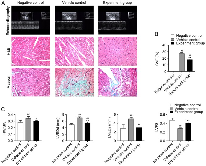
Figure 1.
(A) H&E and Masson's staining in left ventricular tissue slices in age-matched untreated rats, immunized rats treated with vehicle and immunized rats treated with HuMSCs. Magnification, ×100. (B) CVF for Masson's staining in left ventricular tissue slices from the different treatment groups. ##P<0.01 vs. negative control; *P<0.05, **P<0.01 vs. vehicle control, n=8. (C) Heart cavity measurements. ##P<0.01 vs. negative control; *P<0.05, **P<0.01 vs. vehicle control, n=8. H&E, hematoxylin and eosin; HuMSCs, human umbilical cord-derived mesenchymal stem cells; CVF, collagen volume fraction; HW/BW, heart weight/body weight; LVEDd, left ventricular dimension in end diastole; LVEDs, left ventricular dimension in end systole.

