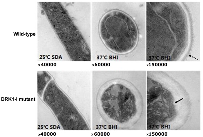
Figure 4.
Hyphal and conidial sections of wild-type and SsDRK1-i strains were observed by transmission electron microscopy. The SsDRK1-i strain displays cell walls with increased and variable thickness. The dashed line arrowhead indicates the electron-dense outer layer of wild-type yeast cells. The black arrowhead indicates the plasma membrane invaginations in the SsDRK1-i conidia. Ss, Sporothrix schenckii; i, interference; SDA, sabouraud dextrose agar; BHI, brain heart infusion.

