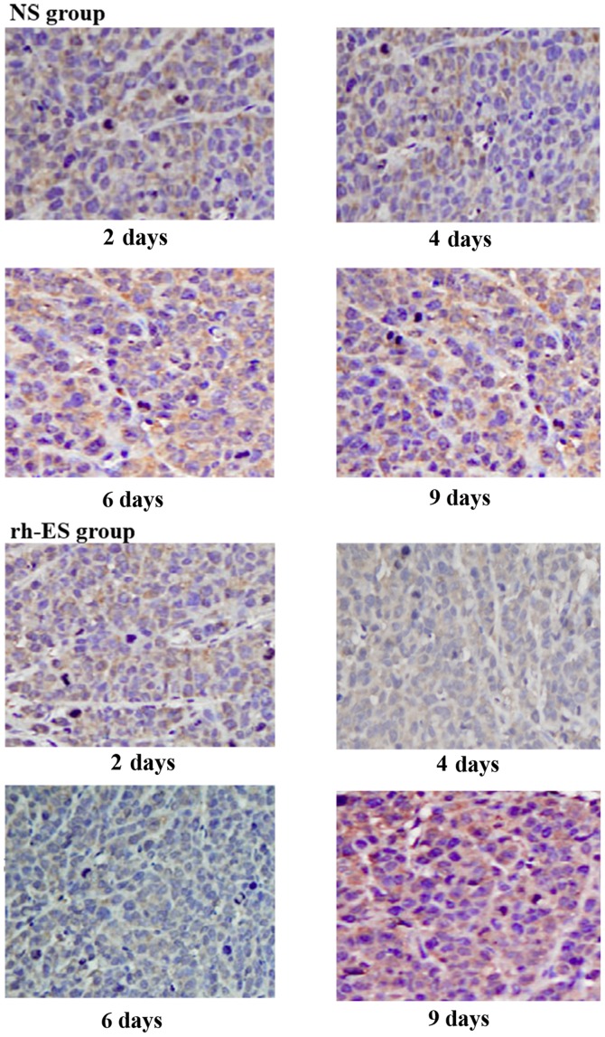
Figure 4.
Representative images showing CA9 (a marker for tumor hypoxia) immunohistochemistry staining in Lewis lung cancer tumors following treatment with rh-ES or NS for days 2, 4, 6 and 9. The positive CA9 signals were located on the cell membrane and had a color of brown. Magnification, ×400. CA9, carbonic anhydrase 9; rh-ES, recombinant human endostatin; NS, normal saline.

