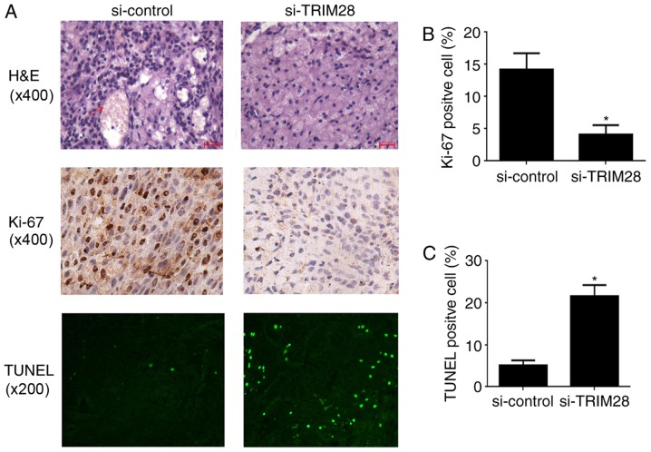
Figure 2.
Histology and immunohistochemistry of tumor tissues from xenografted nude mice. (A) Top, H&E staining. As indicated by the red arrow, the red-stained material in tumors from the control group is erythrocytes and the adjacent structures are blood vessels, which are rarely observed in tumors from PAa/TRIM28-siRNA treated mice (magnification, ×400). Middle, immunostaining for Ki-67 (magnification, ×400). Bottom, TUNEL staining (magnification, ×200). (B) The percentages of Ki-67-positive nuclei was determined on four slides for each group and expressed as mean ± SD. (C) Analysis of the degree of apoptosis in tumors from mice. *P<0.001 compared to the si-control group. H&E, hematoxylin and eosin; si, small interfering; TUNEL, terminal deoxynucleotidyl transferase-mediated deoxyuridine triphosphate nick-end labeling; SD, standard deviation.

