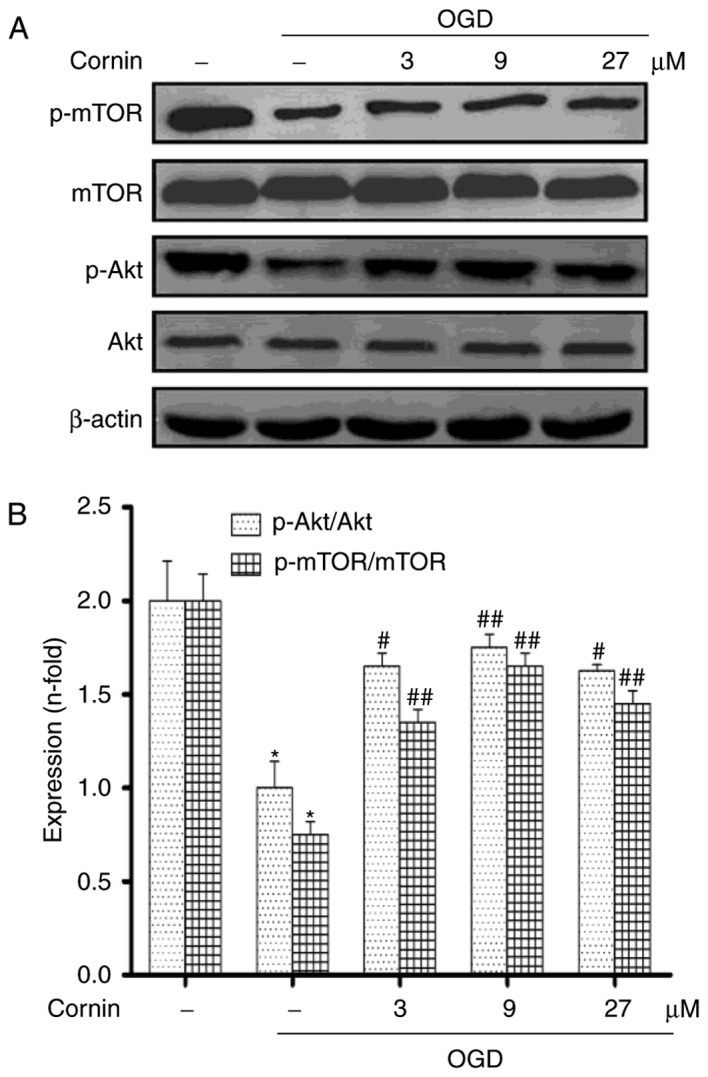
Figure 3.

Effect of cornin on the protein expression of Akt, p-Akt, mTOR and p-mTOR. SH-SY5Y cells were treated with cornin (3, 9 and 27 µM) for 24 h and cell lysates were subjected to immunoblot analysis for detecting the levels of (A) AKT, p-Akt, mTOR, p-mTOR. β-actin was used as the cell lysate loading control. (B) Densitometric analysis was performed. The results are expressed as the mean ± standard deviation. n=3. *P<0.01 vs. control group; #P<0.05, ##P<0.01 vs. OGD group. OGD, oxygen-glucose deprivation; Akt, RAC-α serine/threonine-protein kinase; mTOR, protein kinase mTOR; p, phosphorylated.
