Figure 6.


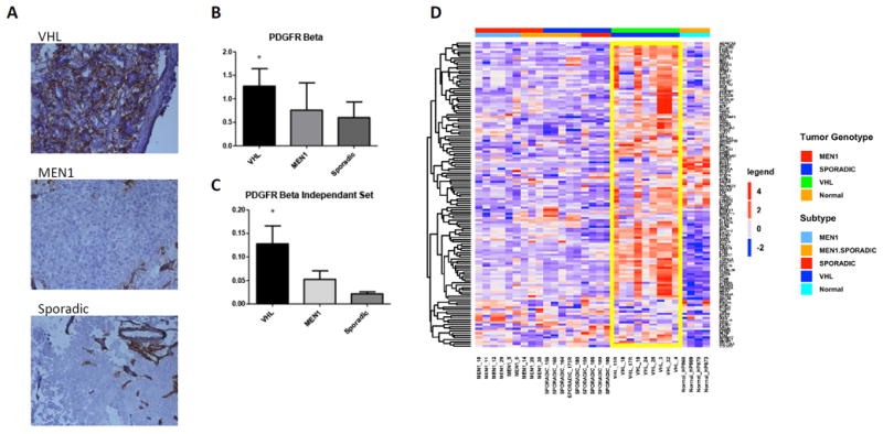
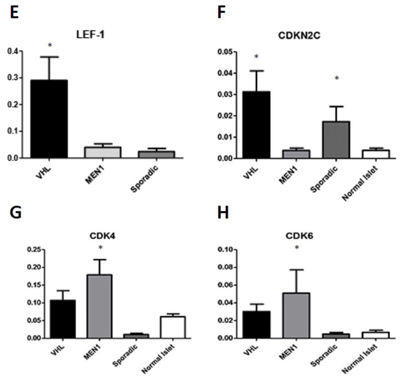
A. Immunohistochemical analysis of PDGFR-Beta in VHL, MEN1 and Sporadic PanNETs shows significantly increased expression in VHL PanNET cells (p<0.05). B. RT-qPCR of PDGFR-Beta confirms microarray analysis and shows significantly increased mRNA levels in the initial cohort. C. Independent validation of PDGFR-Beta expression via RT-qPCR in a separate set of 19 NFPanNETs and four NIC (p<0.05). C. The ESTIMATE (Estimation of Stromal and Immune cells in Malignant Tumours using Expression data) gene signature model confirms increased stromal signature in VHL PanNETs (yellow box). E. RT-qPCR expression analysis showing overexpression of the transcription factor Lef-1 in VHL NFPanNETs when compared to sporadic and MEN1 NFPanNETs. F. RT-qPCR expression analysis showing overexpression the CDK4/CDK6 inhibitor CDKN2C in MEN1 NFPanNETs when compared to sporadic (p=0.06) and VHL NFPanNETs (p=0.02). G. RT-qPCR expression analysis showing overexpression of CDK4 in MEN1 NFPanNETs when compared to sporadic NFPanNETs (p=0.0009) and normal islet cells (p=0.05). H. RT-qPCR expression analysis showing overexpression of CDK6 in MEN1 NFPanNETs when compared to sporadic NFPanNETs (p=0.05).
