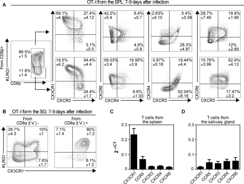
Fig. 7. Most CD8+ T cells in the parenchyma of the salivary gland lack KLRG1 and CX3CR1, but express multiple other chemokine receptors.

Naïve OT-Is (2×10³) were transferred to naïve B6 mice that were infected via the i.p. route one day later with MCMV expressing OVA. Mice were sacrificed 7 or 9 days after infection. (A) Chemokine receptor expression within the KLRG1+ and KLRG1− subsets of the OT-I T cells in the spleen. (B) KLRG1 and CX3CR1 expression of OT-Is from the vasculature (I.V.+) and parenchyma (I.V.−) portions of the salivary gland. In A and B, the data show concatenated FACS plots from one representative experiment at day 9 (n= 3) with mean ± SEM values in each quadrant derived from all experiments (total n=8). (C and D) Naive OT-Is were transferred as in A and sorted from the spleen (B) and the salivary gland (C) 7 days after infection with MCMV expressing OVA. Chemokine receptor expression was assessed on sorted T cells by RT-qPCR. Data were combined from 2-5 independent RT-qPCR assays per sample, with cDNA from OT-I T cells sorted from 2-3 independent mice. Error bars represent SEM.
