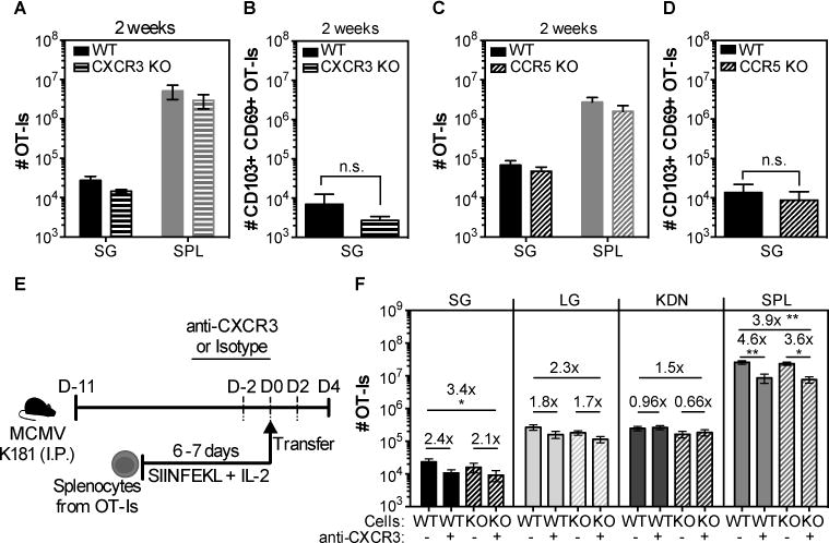
Fig. 8. Lack of CXCR3 or CCR5 does not impact the accumulation of CD8+ T cells in the salivary gland after MCMV infection.

(A and C) Wild-type (WT) OT-Is and OT-Is that lacked either the chemokine receptor CXCR3 or CCR5 were mixed (1×10³ of each) and co-transferred to naïve B6 mice that were then infected with MCMV-Ova. Shown are the overall numbers of CXCR3 KO (A) or CCR5 KO (C) versus wild-type (WT) OT-Is from the spleen (SPL) and the parenchyma of the salivary gland (SG) 14 days after infection. (B and D) Shown are the absolute numbers of CD103+ CD69+ OT-I T cells from the SG. Data are combined data from 2 independent experiments for each KO (dashed bars)/WT (filled bars) pair (n=7 for the WT/CXCR3 and n=5 for the WT/CCR5 experiments). E) Experimental design of the OT-I adoptive transfer for F. WT and CCR5 KO OT-Is were activated in vitro, and 4×10⁶ of each were mixed. Mixed cells were treated with either anti-CXCR3 antibody or an isotype control and co-transferred to MCMV infected mice that were treated with anti-CXCR3 (+) or an isotype control antibody (-) via i.p. injections every other day starting 2 days before the adoptive transfer. F) Shown are the absolute numbers of WT (filled bars) or CCR5 KO (dashed bars) OT-I T cells in the parenchyma of the salivary gland (SG), lungs (LG), kidneys (KDN) and from the overall CD8β+ T cells from the spleen (SPL) of CXCR3 blocked (+) or isotype control-treated (-) mice. Data from 2 independent experiments (n=7 for the isotype treated group and n=8 for the CXCR3 treated group). Error bars represent that SEM. The statistical significance was measured by unpaired t-test after log10 conversion of the absolute numbers (A-D) and One-way ANOVA (F).
