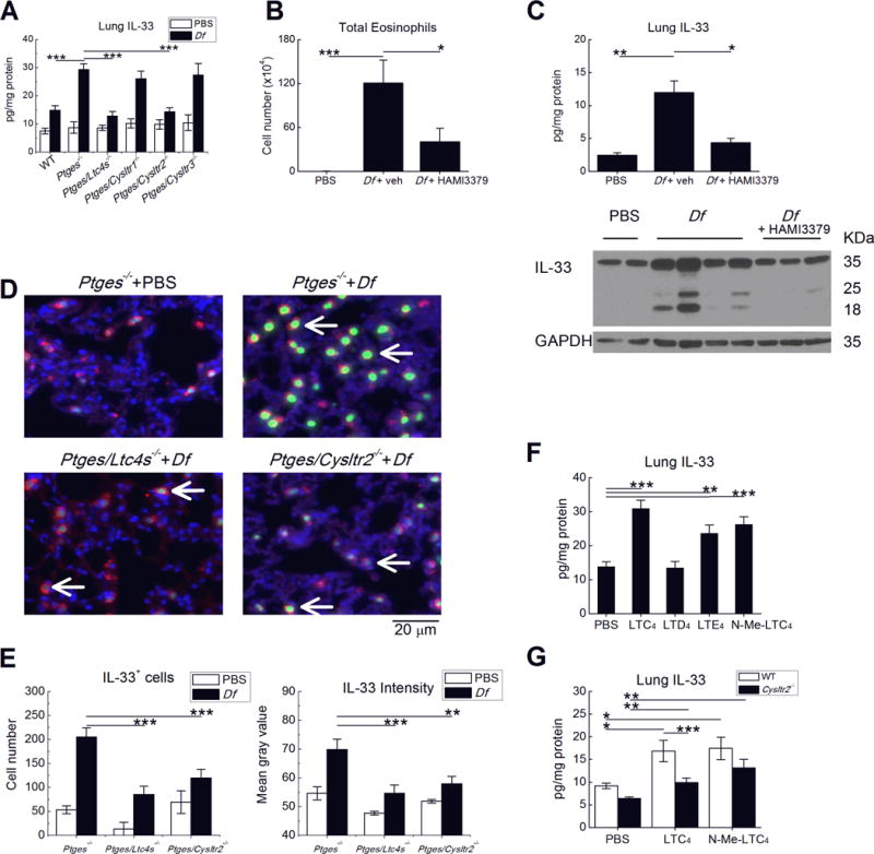
Figure 4. CysLT2R is necessary for IL-33 expression induced by endogenous or exogenous cysLTs.

A. Levels of IL-33 protein detected in the lysates of lungs from the indicated strains of mice treated on six occasions with Df (3 µg) intranasally, or with PBS. B. Total BAL fluid eosinophils and C. Lung IL-33 levels (top) in Ptges−/− mice treated daily with the CysLT2R-selective antagonist HAMI-3379 during priming with Df. Western blot (bottom) showing full length (~35 kDa) and cleaved forms of IL-33 in whole lung lysates from Ptges−/− mice of the indicated treatment groups. D. Immunofluorescent stains for IL-33 (green) localizing to the nuclei (blue) of SPC (red)-expressing lung AT2 cells from representative mice of the indicated strains and treatment groups. E. Quantitative analysis of numbers (left) and per cell intensity (right) of IL-33 staining in AT2 cells. F. Effects of exogenous cysLTs on lung IL-33 protein in OVA-sensitized and challenged PGE2 sufficient mice. G. Effect of CysLT2R deletion on lung IL-33 expression induced by the indicated exogenous cysLTs. Results in A-C and E-G are from 10 mice/group. *** P < 0.001, ** P < 0.01, * P <0.05.
