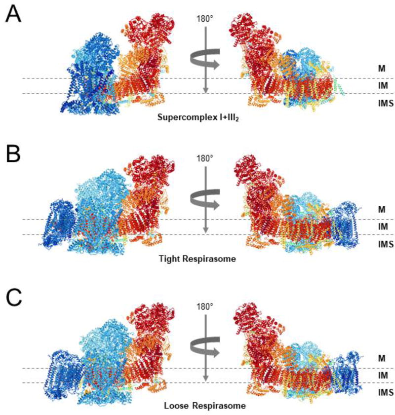
Figure 1. Architectures of the mammalian respirasomes.
Side views along the membrane from (A) Supercomplex I+III2, (B) the tight respirasome, and (C) the loose respirasome, according to the structures proposed by Letts et al. [22]. Images were obtained from the RCSB Protein Data Bank in combination with the NGL viewer. The structural models of CI, CIII2, and CIV are colored in red, turquoise, and navy blue, respectively. The transmembrane region is indicated by two dashed lines. M, matrix; IM, mitochondrial inner membrane; IMS, intermembrane space.

