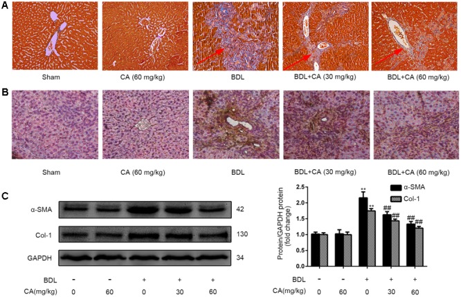
FIGURE 2.

Carnosic acid significantly ameliorates BDL-induced liver fibrosis in rats. (A) Masson staining for collagen deposition in rat liver sections (x200). The hyperplasia of the lattice fibers and collagenous fibers has been marked with arrows. (B) Immunohistochemistry for α-SMA in rat livers (x200). (C) The protein expression of α-SMA and type 1 collagen in the liver was measured by western blot (n = 3); the results were normalized relative to the expression of GAPDH. The data are presented as the means ± SD. ∗∗P < 0.01 versus the sham group; ##P < 0.01 versus the BDL group.
